Echographie gynécologique

A black and white photograph of a pregnant person holding an ultrasound image in front of a polka dot dress. Hands gently cradle the belly, conveying a sense of care and anticipation.

Échographie de contrôle de vacuité utérine (post-fausse couche ou IVG)

A person wearing blue medical scrubs and gloves operates an ultrasound machine in a clinical setting. The individual's hands are adjusting the controls and handling the transducer probe, which is resting on the console equipped with various buttons and dials.

Échographie de contrôle de stérilet (DIU)

A person wearing blue medical scrubs and gloves operates an ultrasound machine in a clinical setting. The individual's hands are adjusting the controls and handling the transducer probe, which is resting on the console equipped with various buttons and dials.

Échographie pelvienne (contrôle kystes, douleurs pelviennes)

Echographie pour s'assurer que tout va bien après une fausse couche ou une interruption de grossesse.

Echographie pour s'assurer que le stérilet est bien positionné et qu'il remplit son rôle.

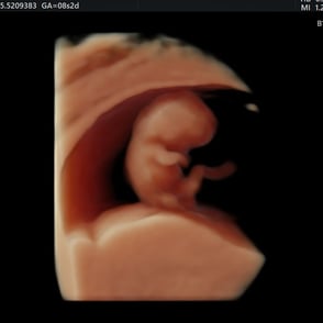
Échographie de Datation

Echographie de datation permet de dater la grossesse en mesurant la taille de l'embryon, de vérifier son développement et de confirmer que la grossesse se déroule au bon endroit (dans l'utérus).

Échographies obstétricales (1er au 3ème trimestre)

Échographie de contrôle de croissance

Echographie obstétricale

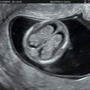
L'échographie du 1er trimestre (entre 12 à 13 SA) permet de dater de façon fiable la grossesse (+/- 5 jours), vérifier la bonne évolutivité de celle-ci, dépister de potentielles anomalies, et de mesurer la clarté nucale (paramètre qui rentre en compte dans le dépistage de la trisomie 21).

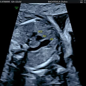
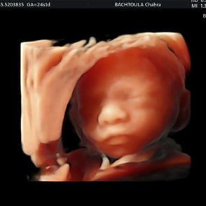
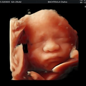
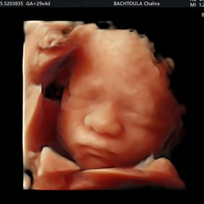
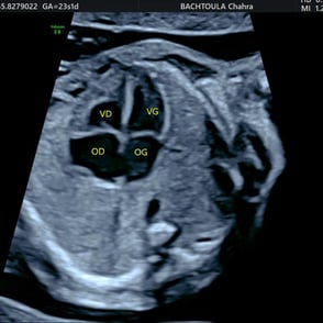
L'échographie du 2e trimestre (entre 22 et 24 SA) est la principale échographie permettant d'analyser la morphologie foetale, dépister certaines anomalies morphologiques, estimer le poids et la bonne croissance du bébé ainsi que d'autres paramètres importants (position du placenta, la quantité de liquide amniotique, les dopplers uterins et ombilical).

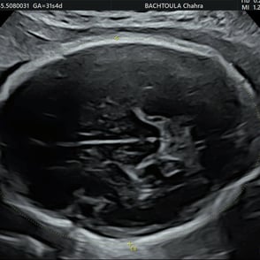
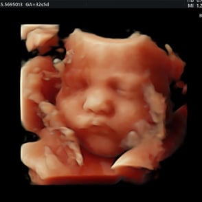
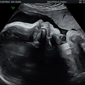
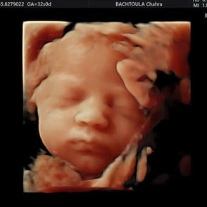
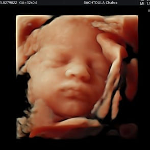
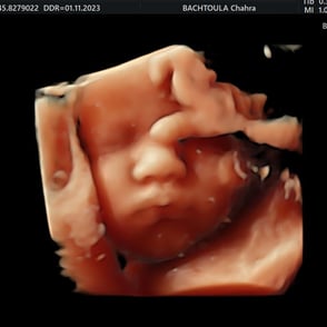
L'échographie du 3e trimestre (entre 32 et 34 SA) nous permet de refaire un examen morphologique du bébé, d'apprécier sa croissance et de vérifier sa position avant l'accouchement.

A person wearing blue medical scrubs and gloves operates an ultrasound machine in a clinical setting. The individual's hands are adjusting the controls and handling the transducer probe, which is resting on the console equipped with various buttons and dials.

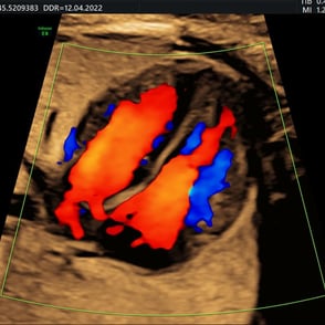
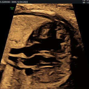
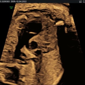
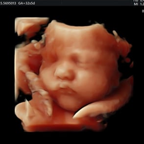
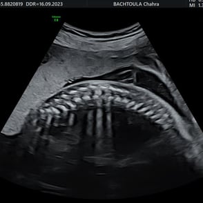
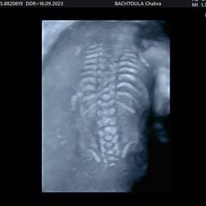
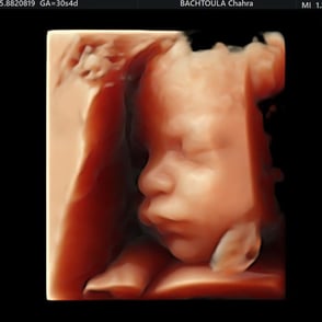
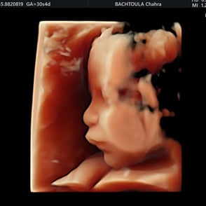
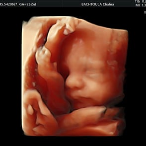
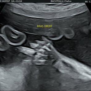
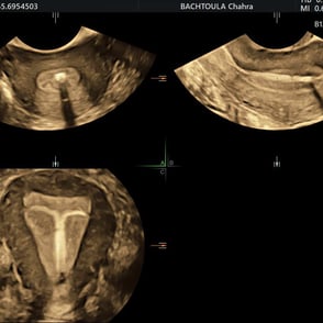
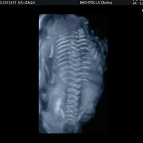
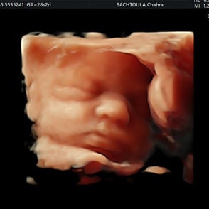
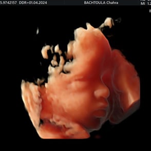
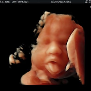
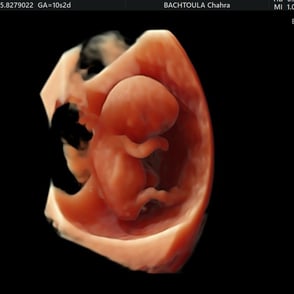
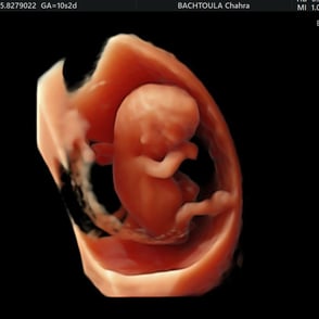
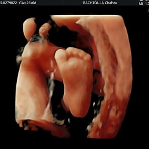
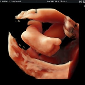
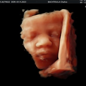
Galerie des clichés d'échographie